Saturday, September 8, 2012

Functional Programming


Today I was hacking on my TDT reading code when I came across the following ugly code:
if map(iscomplex, map(np.squeeze, map(np.array, args))):
    # ...
I thought to myself, "Hey why not throw some functional programming at this?", to get
if composemap(iscomplex, np.squeeze, np.array)(args):
    # ...
Definitely more readable if a little too terse.

What's great is that I can pass an arbitrary number of functions to compose map and it will run each function on all the arguments in args. Sweet.

How is it actually implemented?
def compose2(f, g):
    if not all(map(callable, (f, g))):
        raise TypeError('f and g must both be callable')
    return lambda *args, **kwargs: f(g(*args, **kwargs))
This little gem just returns a function that calls f and g with any number of arguments. Totally jacked from the Python functools module docs.
from functools import partial, reduce
from itertools import repeat

def composemap(*args):
    partial_map = map(partial, repeat(map, len(args)), args)
    return reduce(compose2, partial_map)
That's it! Now, to be sure there's a ton of stuff going on here and it took me a while to grok it, so let's break it down line by line.

The partial function takes a callable and optional positional and keyword arguments and returns another callable that has the given arguments already filled in. It's similar to currying, but a little different.

The first line of composemap results in
[partial(map, arg0), partial(map, arg1), ... partial(map, argN)].

This makes sense because what I want to do is actually run map, but with the functions I've provided as arguments first and whatever sequence i decided to map each of the functions I've provided over. However, I don't want to evaluate this right away so I create a list (actually a tuple) that contains all of the partial applications that I want to perform when I provide a sequence to map over.

OK whew! That was a lot to take in.

For the last line recall what partial does: it takes a callable with part of the sequence arguments that you want to provide and returns a callable with those arguments already set to go when you call the function with the final arguments (or more arguments).

Look at the reduce documentation for more details on reduce. If you're familiar with Haskell you can think of reduce as the Python version of foldl.

What I've done here is give reduce the list of partial functions that I want to eventually evaluate.

What the end result looks like is something like this

Given a function $h = f\circ g$ and an $n$-sequence of partial functions $\mathbf{p} = \left(p_1, p_2,\ldots,p_n\right)$ then the "reduce" function, in this case is given by
\begin{equation}
\textrm{reduce}\left(h, \mathbf{p}\right) = h\left(\ldots h\left(p_1, p_2\right), \ldots p_n\right)
\end{equation}

Recall that $h\left(f,g\right)$ returns a function. So running $h$ with $p_1,p_2$ as arguments gives $p_0\left(p_1\left(\cdot\right)\right)$. Pass that into $h$ with the third element of $\mathbf{p}$, $p_3$. Keep doing that until all of the arguments are eaten up.




Sunday, February 26, 2012

Definition of a Confidence Interval

"95% of intervals of the form $$\left({\overline X} - \frac{2\sigma}{\sqrt{n}},{\overline X} + \frac{2\sigma}{\sqrt{n}}\right),$$ which are based on samples of size $n$ each, will contain the mean of the normal density in their interior."

Saturday, February 11, 2012

Convexity

Some things to prove:
  1. The distance to a set $S$ from some vector $\mathbf{x}$, given by $d\left(\mathbf{x}, S\right) = \displaystyle\min_{\mathbf{y}\in S}\,\,\|\mathbf{x} - \mathbf{y}\|$ is convex.
  2. The set of of all symmetric positive definite matrices is convex.
  3. Let $f\left(x\right)=\exp\left(Q\left(x\right)\right)$. Prove that $f$ is log concave if $Q\left(x\right)$ is concave.

To do this, I'm going to employ the use of some tools from Chapter 2 of the book Convex Optimization, by Steven Boyd and Lieven Vandenberghe. One of the great things about this book is that it is very accessible.

First, recall the definition of lines and line segments: If two points $x,y$ in $\mathbb{R}^{n}$ are not equal, then points of the form the form $y = \theta x_1 + \left(1 - \theta\right)x_2$ where $\theta \in \mathbb{R}$ form the line passing through $x_1$ and $x_2$.

This is nice and all, but it's not exactly the most intuitive way to define a line. The authors graciously give another interpretation which I find to be very intuitive: $$y=x_2+\theta\left(x_1-x_2\right).$$ They explain it in an equally intuitive way: "... $y$ is the sum of the base point $x_2$ (corresponding to $\theta = 0$) and the direction $x_1 - x_2$ (which points from $x_2$ to $x_1$) scaled by the parameter $\theta$." (p. 21)

Let's look at this in vector notation. $y$ should actually be $\mathbf{y}$ because we're not talking about scalar values here, we're talking about points/vectors on the $n$-dimensional real line, which have more than one component. The same goes for $x_1$ and $x_2$. So, it should be written $$\mathbf{y} = \mathbf{x}_2 + \theta\left(\mathbf{x}_1 - \mathbf{x}_2\right).$$ It's one of my math pet peeves when people don't use something to indicate whether they are talking about vectors or scalars, even if you define it that way from the start. We're so used to working with symbols like $x$ and $y$ as scalar values, that it's nice to be able to let the paper take care of the notation and not have to think "Am I looking at a scalar or vector here?". When vectors are bold you can immediately see that the data type is a vector, whereas with the other convention $x$ and $y$ could be a scalar or a vector depending on how it was defined at the beginning of the [proof, book, paper, etc.]. OK, rant over!

Assuming you have NumPy and matplotlib installed you can use the following Python code to demonstrate the definition of a line i just described.
from pylab import *

def randbetween(a, b, *size):
    assert a < b, 'a must be less than b'
    for s in size:
        assert isinstance(s, int), \ 
            'the elements of size must all be integers'
    bma = b - a
    return bma * rand(*size) + a

b = 10
a = 1
x1 = randbetween(a, b, 1, 2)
x2 = randbetween(a, b, 1, 2)

# 0 <= theta <= 1
theta = linspace(0, 1, 10000)[newaxis]

# compute the line over theta
y = dot(theta.T, x1) + dot(1 - theta.T, x2)

# plot the points and the line
figure()
hold('on')
plot(x1[0, 0], x1[0, 1], '.', ms=20)
plot(x2[0, 0], x2[0, 1], '.', ms=20)
plot(y[:, 0], y[:, 1])
hold('off')

# show the plots
show()
You can literally just copy this code, then in an IPython console type: paste. This will run all of the code I just listed and should result in a nice plot. Obviously for $\theta > 1$ and $\theta < 0$ the line will extend beyond $\mathbf{x}_1$ and beyond $\mathbf{x}_2$ respectively. What's more, you can rewrite the second equation in vector-matrix form. Given two $k$-dimensional row vectors $\mathbf{x}_1$ and $\mathbf{x}_2$ and given a column vector $\boldsymbol\theta = \left(\theta_1, \theta_2, \dotsc, \theta_n\right)$ where $\theta_1 = 0$ and $\theta_n = 1$ and monotonically increasing elements, then a line in $\mathbb{R}^{n}$ can be written as $$\mathbf{Y} = \boldsymbol\theta\cdot\mathbf{x}_1 + \left(\mathbf{1}_n - \boldsymbol\theta\right)\cdot\mathbf{x}_2,$$ where $\mathbf{1}_n$ is an $n\times{1}$ vector with each element equal to 1, and $\mathbf{Y}$ is an $n\times{k}$ matrix with each column being the points of the line in the $k$th dimension. The next important concept is the notion of an affine set. A set $C\subseteq{\mathbb{R}^{n}}$ is said to be affine if the line through any points in $C$ not equal to each other is in $C$. Said differently, "$C$ contains the linear combinations of any two points in $C$, provided the coefficients in the linear combination sum to one (Boyd & Vandenberghe)." (p. 22). In symbolic terms, $$x_1, x_2\in{C}\textrm{ and }\theta\in{\mathbb{R}}\Rightarrow{\theta{x_1}+\left(1-\theta\right)x_2\in{C}}$$ OK, now on to convex sets. The definition is exactly the same as an affine set except that $\theta$ is bounded by 0 and 1. A set $C$ is convex if, $$x_1,x_2\in{C}\textrm{ and }\theta\in\left[0,1\right]\Rightarrow{\theta{x_1}+\left(1-\theta\right)x_2\in{C}}$$ Let's now define a convex function. A function $f:\mathbb{R}^{n}\rightarrow\mathbb{R}$ is said to be convex if the domain of $f$ is a convex set and if for all $x$, $y$ is an element of the domain of $f$, and $\theta$ with $0\leq\theta\leq{1}$ we have, $$f\left(\theta x + \left(1-\theta\right)y\right)\leq\theta f\left(x\right) + \left(1-\theta\right)f\left(y\right).$$ Strict convexity is when $x\neq{y}$ and $0\lt\theta\lt{1}$ and concavity is when $-f$ is convex, with strict concavity if $-f$ is strictly convex.

Let's use the definition of convexity to show that $d$ is convex.

The triangle inequality and homogeneity show us that $$f\left(\theta\mathbf{x} + \left(1-\theta\right)\mathbf{y}\right)\leq f\left(\theta\mathbf{x}\right) + f\left(\left(1-\theta\right)\mathbf{y}\right)=\theta f\left(\mathbf{x}\right) + \left(1-\theta\right)f\left(\mathbf{y}\right)$$ is true. Then substituting $d$ for $f$ allows us to see that $d$ is convex.

In my next few posts I'll show the 2 and 3 parts of the list at the beginning of this post.

Uninstall All Ruby Gems

Today I ran into an issue when trying to do a nice Rails tutorial. Having two versions of Ruby installed on my machine was (I think) causing some conflicts. At best, it was confusing me. So, here's how to uninstall all Ruby gems:

gem list | cut -d" " -f1 | xargs gem uninstall -aIx

Tuesday, February 7, 2012

Gamma

I was working on some mathematical statistics problems and I came across a slick estimation problem that I thought I would share.

Problem:
Given $$\left(f\mid k\right) = \frac{x^{k-1}\exp\left(-x/\theta\right)}{\Gamma\left(k\right)\cdot\theta^{k}},\,\,\,\textrm{ where } x \gt 0,\,\,\, k \gt 0,\,\,\, \theta = 1,$$Determine whether it is possible to find a value of $c$ such that $d\left(X\right) = cX^{2}$ will be an unbiased estimator of $k$.

My attempt at a solution:

$$E\left[c\cdot X^{2}\right] = k$$ for the left hand side to be an unbiased estimator of $k$. $$c\cdot E\left[X^{2}\right] = k$$ is true via the properties of $E\left[\cdot\right]$. Rearranging terms we can see that $$c = \frac{k}{E\left[X^{2}\right]}$$ $$c = k\cdot\left(\int_{0}^{\infty}x^{2}\frac{x^{k-1}e^{-x}}{\Gamma\left(k\right)}\mathrm{d}\,\,x\right)^{-1} = k\cdot\left(\int_{0}^{\infty}\frac{x^{k+1}e^{-x}}{\Gamma\left(k\right)}\mathrm{d}\,\,x\right)^{-1}$$ $$k\cdot\Gamma\left(k\right)\cdot\left(\int_{0}^{\infty}x^{k+1}e^{-x}\mathrm{d}\,\,x\right)^{-1} = \frac{\Gamma\left(k+1\right)}{k\cdot\Gamma\left(k+1\right)}=\frac{1}{k}.$$ So the value of $c$ such that $E\left[cX^{2}\right]$ is NOT an unbiased estimator of $k$ is $1/k$ because by definition, an unbiased estimator of some parameter $\theta$ cannot depend on the value of $\theta$.

Saturday, February 4, 2012

Setting up Git for my website

Just wanted to log the process of setting up the Git version control system for editing the code of my website.

  • Local Machine
    • Make a directory for your code
    • Run git init inside of it
    • Add some content and commit it
  • Server
    • Password stuff
      • Make an SSH public encryption key if you haven't already
        • ssh -t rsa -C 'someone@somewebsite.com'
        • Follow the instructions it spits out
        • Give a password if you want
      • ssh -p 2222 me@myserver.com 'mkdir -p .ssh'
        • You'll have to enter a password here
      • cat ~/.ssh/id_rsa.pub | ssh -p 2222 me@myserver.com 'cat >> .ssh/authorized_keys'
    • Git stuff
      • ssh -p 2222 me@myserver.com
      • cd public_html && mkdir website.git && cd website.git && git init --bare
      • cd hooks && cp post-receive.sample post-receive
      • echo 'GIT_WORK_TREE=/dir/where/you/want/to/store/code git checkout -f' >> post-receive
  • Local Machine
    • git remote add website ssh://ip.add.re.ss:portnumber/~/public_html/website.git
    • git push -u website +master:refs/heads/master && git push
Now, if I want to edit the code of my website on my local machine I can do so and have changes pushed to the website whenever I want to. I just have to be careful not to push breaking changes. Time to get intimate with Git branches!

Funny

Saw this picture while trolling some links from Hacker News.

#4 reminds me of some arguments I've had...

Wednesday, February 1, 2012

Advanced Topics in Statistics: Day 2

Time for some more probability review and some new material on classes of distributions. Chapter numbers refer to Casella and Berger (2002).

More review of probability:
2.2:  Expected Values: *Very Important*
$$E\left[g\left(X\right)\right] = \int_{-\infty}^{\infty}g\left(x\right)f_{X}\left(x\right)\,\,\mathrm{d}x,$$ when $X$ is a continuous random variable.
$$E\left[g\left(X\right)\right] = \sum_{x}g\left(x\right)f_{X}\left(x\right),$$ when $X$ is a discrete random variable.
Properties of $E\left[\cdot\right]$:
  • $E\left[a\right] = a, a\in\mathbb{R}$
  • $E\left[ag_{1}\left(X_{1}\right) + bg_{2}\left(X_{2}\right) + c\right] = aE\left[g_{1}\left(X_{1}\right)\right] + bE\left[g_{2}\left(X_{2}\right)\right] + c$
  • If $g_{1}\left(x\right)\le g_{2}\left(x\right)\le \cdots \le g_{n}\left(x\right), \forall x,$ then $E\left[g_{1}\left(x\right)\right]\le E\left[g_{2}\left(x\right)\right]\le\cdots\le E\left[g_{n}\left(x\right)\right]$.

Remark I
In measure theoretic notation the generalized expected value is, $$E\left[x\right] = \int_{\omega\in\Omega}X\left(\omega\right)\,\,\mathrm{d}P\,\left(\omega\right).$$ I'm not sure exactly what this means; I'll have to come back to it later.

Remark II
What about interchanging sums and integrals?$\newcommand{\?}{\stackrel{?}{=}}$
$$\begin{eqnarray*}
\int\int f\left(x,y\right)\,\,\mathrm{d}x\,\mathrm{d}y&\?&\int\int f\left(x,y\right)\,\,\mathrm{d}y\,\,\mathrm{d}x\\
\sum_{j}\sum_{k}a_{jk}&\?&\sum_{k}\sum_{j}a_{jk}\\
\sum_{j}\left(\int f_{j}\left(x\right)\,\,\mathrm{d}x\right) &\?& \int\left(\sum_{j}f_{j}\left(x\right)\right)\,\,\mathrm{d}x
\end{eqnarray*}$$
For example, $a \ge 0, f \ge 0, f_{j} \ge 0$ then it works. Or, take the absolute values inside the sum (integral), and show that one of the sides is finite.

More to follow on classes of distributions.
Here's how to mount and unmount an ISO image in Linux, from Bash.

Mounting:

sudo mkdir /media/example
sudo mount -o loop example.iso /media/example


Unmounting:

sudo umount /media/example
sudo rmdir /media/example

Basic Probability

Approximately 1/3 of all human twins are identical (one-egg) and 2/3 are fraternal (two-egg) twins. Identical twins are necessarily the same sex, with male and female being equally likely. Among fraternal twins, approximately one-fourth are both female, one-fourth are both male, and half are one male and one female. Finally, among all U.S. births, approximately 1 in 90 is a twin birth. Define the following events:

$A = \left\{\textrm{a U.S. birth results in twin females}\right\}$
$B = \left\{\textrm{a U.S. birth results in identical twins}\right\}$
$C = \left\{\textrm{a U.S. birth results in twins}\right\}$

(a) State in words, the event $A\cap{B}\cap{C}$.

  • A U.S. birth results in identical twin females.

(b) Find $P\,\,\left(\!A\cap{B}\cap{C}\,\right)$.

  • $P\,\,\left(\!A\cap{B}\cap{C}\,\right) = \frac{1}{90}\cdot \frac{1}{3}\cdot \frac{1}{2}=\frac{1}{540}$

Saturday, January 28, 2012

Neuroanatomy: End

I finished up the last component of my neuroanatomy course today.

This was one of the more intense courses I've taken during my Ph.D, simply because we had two weeks to learn almost every aspect of neuroanatomy as was humanly possible. For some reason courses like this one seem to bring out my philosophical side; maybe it's because in this case it was such a graphic depiction of this blob in our heads that we so often take for granted.

It's amazing to see the brain in all of its glory. Slicing structures left, right, top, bottom, and sideways really gives you an idea of how complicated this hunk of wet machinery really is.

Tuesday, January 24, 2012

Dynamic Reorganization of striatal circuits during the acquisition and consolidation of a skill

  • Authors: Yin, Mulcare, Hilario, Clouse, Holloway, Davis, Hansson, Movinger, Costa
  • Summary: 
    • Region- and pathway specific plasticity sculpts the circuits involved in the performance of the skill as it becomes automatized.
  • Background: 
    • Dorsomedial (associative; caudate in primates) striatum (DMS) receives input primarily from association cortices such as the prefrontal cortex (PFC). The dorsolateral (sensorimotor; putamen in primates) striatum (DLS) receives input from sensorimotor cortices is critical for the more gradual acquisition of habitual and automatic behavior.
    • CPC: 
      • What role does the cerebellum play, if any, in the process of "transferring" newly learned skills to some sort of storage? Is it too general to say that DMS is involved in learning causal relationships, whereas DLS is involved in automatic association of these causes and effects?
  • Question(s): 
    • Can changes in striatal neural activity observed during skill learning be mediated by synaptic plasticity or excitability chagnes in medium spiny projections neurons in the dorsal striatum using an ex vivo (a slice preparation) approach.
  • Result(s): 
    • "... task-related activity in these striatal regions differed during the acquisition and consolidation of a new skill, with the DMS being engaged during the early phase and the DLS being engaged during the late phase."
    • "[in the ex vivo preparation, ] [l]earning was accompanied by long-lasting changes in glutamatergic transmission."
      • "These changes evolved dynamically during the different phases of skill learning;
      • "Changes in the DMS were predominant early in training, whereas changes in the DLS emerged only after extensive training."
      • "Long-lasting changes in the DLS after extensive training were pathway specific and occurred predominantly in DA receptor 2 (D2)-expressing striatopalidal medium spiny neurons."
  • Conclusion(s):
    • "..., the performance of the skill after extended training became less dependent on the activation of D1-type DA receptors, which are mainly expressed in striatonigral neurons."
  • Method(s):
    • DMS/DLS Involvement in Early and Late Skill Learning
      • Quantified the modulation of activity using the following equation
      • In the DMS the rate modulation increased during the early phase of training but returned to naive levels with further training
      • In the DLS, the modulation of firing rate increased gradually with training.
        • Accompanied by a decrease in baseline firing rate during the intertrial period.
          • The authors interpret this as an increase in signal-to-noise ratio after extended training.
    • Next question:
      • Are the region-specific changes observed in vivo driven by glutamatergic synaptic plasticity or excitability changes in the striatum by training the mice for either 1 day or 8 days on an accelerating rotarod?
      • This was followed up by examining ex vivo the changes in DMS and DLS related to the different phases of skill learning.

Functional architecture of basal ganglia circuits: neural substrates of parallel processing

Authors: Garret E. Alexander, Michael D. Crutcher
Summary: This paper discusses the evidence for parallelization of circuitry within the individual circuits of the basal ganglia.


  • Previous Views:
    • The basal ganglia served to integrate converging influences from cortical association and sensorimotor areas during their passage through basal ganglia to common thalamic target zones.
    • Same BG recipient zones in thalamus received ascending, convergent inputs from the cerebellum and returned their own projections exclusively to primary motor cortex.
  • More Recent Evidence:
    • BG and cerebellar projections are directed to completely separate target zones with the thalamus.
    • Association and sensorimotor cortex form closed circuits between cortex, BG, and thalamus.
      • Output projects not only to primary motor cortex, but to virtually the entire frontal lobe.
  • Main Question: 
    • Is a parallel functional architecture evident within individual basal ganglia circuits?
  • Motor Circuit:
    • Features Intrinsic to Parallel Organization
      • Cortex $\rightarrow$ glutamatergic $\rightarrow$ striatum $==$ input
      • Basal Ganglia $\rightarrow$ tonic, GABA-mediated inhibitory effect on nuclei in thalamus
        • modulated by 2 opposing but parallel pathways that pass from striatum $\rightarrow$ basal ganglia output nuclei
      • Direct pathway $\rightarrow$ output nuclei
        • comes from inhibitory striatal efferents that contain GABA and substance P $\rightarrow \not$ thalamic stage
      • Indirect pathway $\rightarrow$ GPe $\rightarrow$ subthalamic nucleus via GABAergic pathway $\rightarrow$ excitatory (Glu)
      • GPe neurons exert a tonic inhibitory influence on the subthalamic nucleus.
      • GABA/enk projection from striatum suppress activity of GPe neurons $\rightarrow$ disinhibits the subthalamic nucleus $\rightarrow$ excitatory drive on the output nuclei $\rightarrow$ increases inhibition of their efferent targets within the thalamus.

Monday, January 9, 2012

Neuroanatomy: Day 0

I'm taking an intensive 2-week neuroanatomy course at NYU for the next two weeks. I'll be posting notes and interesting tidbits about things that I'm learning. This post will be on the topic(s) of the first 3 chapters of our textbook.

Neurodevelopment happens to be something that I'm particularly weak on, and the first chapter of our text jumps right into it, for better or for worse.

A section titled: "A note on times and ages" is in part this inspiration for this post. This section points out some very basic clinical conventions used to delimit certain periods of neural development:

1. Pregnancy is timed from the 1st day of the last menstrual period.
2. An 8-week old ball of cells is called a fetus.
3. The embryonic period is divided in to 23 Carnegie stages (not sure where Carnegie comes from), with the neural folds appearing at stage 8.

More on development:
By the end of the 3rd week the neural folds have begun to fuse with one another to form the neural tube. The cells that line the tube will eventually become the neurons that make up all of the neurons in the CNS.


Neural crests are what give rise to many neuroglia and other cells outside of the nervous system. These cells migrate extensively. 

An interesting fact of the brain is that during development there are many more neurons formed than will go on to exist when the animal is an adult. Evidence has shown that in vertebrates cells that died were those that failed to make synaptic connections. Unfortunately, there is currently no evidence to suggest that adult human brains regenerate neurons (with the exception of an area of the hippocampus and olfactory neurons).

The developing nervous system is conventionally divided into 5 parts and their names change as the organism develops:
  1. Telencephalon $\rightarrow$ Cortex
  2. Diencephalon $\rightarrow$ Thalamus/Hypothalamus
  3. Mesencephalon $\rightarrow$ Midbrain
  4. Metencephalon $\rightarrow$ Pons and Cerebellum
  5. Myelencephalon $\rightarrow$ Medulla Oblongata
Here's a nice breakdown from the text:


Another interesting part of neural development is the construction of the meninges. The meninges are a membraneous tissue that protect the brain. The meninges is somewhat like plastic wrap for the brain, at least upon initial visual inspection.

Key parts of the brain:

  • Spinal cord
    • Least differentiated part of the nervous system
    • pair of nerves is connected to the spinal cord (dorsal sensory root, ventral motor root)
    • H-shaped spatial organization with gray matter as the H and white matter in the surrounding area
      • Gray matter in the spinal cord is important for reflexes
  • Medulla oblongata
    • A continuation of the spinal cord
  • Pons
    • Two-part structure
      • Tegmentum
        • ascending/descending tracts
      • Basal pons
        • Provides connections between the cortex of a cerebral hemisphere and that of the contralateral cerebellar hemisphere
  • Midbrain
    • Dorsal region
      • Tectum: lower level visual/auditory control
      • Red nucleus/substantia nigra: important in motor control
      • attached to the mid brain via the superior cerebellar peduncles
  • Cerebellum
    • Extremely large, contains most of the neurons of the brain
    • Important for muscular compensation during movement
  • Diencephalon
    • Central core of the cerebrum
    • Largest component is the thalamus
    • Epithalamus and pineal gland constitute an endocrine organ
    • Interestingly, the retina is a derivative of the diencephalon
  • Telencephalon
A human brain (!) :


Monday, November 14, 2011

Declarative Memory

This is going to be quite a long post because we've got four articles to read for this week's lecture. The topic is declarative memory.

Article: A unified framework for the functional organization of the medial temporal lobes and the phenomenology of episodic memory
Author: Charan Ranganath


  • Abstract
    • What is the role of the medial temporal lobe in recognition memory (I'll get to what that is in a second)?
    • Evidence is consistent with the notion that MTL subregions differ in terms of the kind of information that they process and represent.
    • These subregions support episodic memory by binding item and context information.
    • My comments
      • What kind of information are we talking about here (I'm asking this question in the most concrete sense, with 'kind' here meaning some physically realizable data structure, not 'Oh, it "represents" short-term memories')?
      • How is this information processed (i.e., what (neuronal) procedures (computations) underlie such processes)?
      • What and how is this information represented?
      • Side rant
        • I'm so annoyed by neuroscientists throwing the words information, process, representation around as if they actually mean something when they say it. I feel like it is used to cover up ignorance about whatever said person is talking about.
        • Presumably, we're talking about encoding, but no one actually knows how the brain encodes sensory input (e.g., how does the brain transform light into the color red?). That is, there's no indication as to what the brain's encoding scheme is other than that it probably involves spike trains.
      • What in the hell does it mean to bind item and context information?
        • Are they being 'added' together in some sense? Multiplied? Composed?
          • I'm going to go out on a limb and suggest that the author didn't really consider what binary operation that neurons are carrying out to cause this so-called binding to occur.
  • History of Recognition Memory
    • Milner and colleagues showed that extensive damage to the MTL impairs the formation of new memories for events, while sparing many other processes, such as motor skill learning.
    • Research in monkeys corroborates this finding, especially on a delayed non-match to sample task with a long delay or large list of items to-be-remembered is given.
    • More research suggested that the perirhinal cortex (PRc) plays a large role in recognition memory (same task as previous point)
      • Deficits were found even with no delay
      • Lesions of hippocampus proper (or damage to HC through the fornix) had very mild effects on recognition memory
        • Similar effects found in rats and humans
  • Two Component Models of the MTL
    • Cohen et. al's Relational Memory Theory
      • PRc and parahippocampal cortex (PHc) encode specific constituent elements of an event (items)
      • Hippocampus encodes representations of the relationships of the items.
    • Complementary Learning Systems (CLS)
      • Hippocampus is specialized to rapidly encode new information
        • Sparse, minimally overlapping representations that are well suited to encode specific episodes
      • PHc
        • Represents stimuli and events in terms of their constituent features; stimuli with overlapping representations have overlapping neural representations.
    • Aggleton and Brown's Unnamed Model
      • The hippocampus supports recall or recognition based on conscious recollection.
      • HC and PHc differentially support different subjective experiences.
    • What do these models predict about recognition memory?
      • Relational Theory
        • Relational information depends on HC, but PRc should be sufficient to support item recognition without relational information
      • CLS
        • Emphasizes the distinction in the processes that underlie recognition memory (familiarity vs. recollection).
          • What is the degree of overlap in recognition memory signals elicited by studied items and unstudied lures?
        • The cortical component of the model produces a signal that indexes the global match between a cue and all previously learned information due to broad overlapping stimulus representations.
        • The hippocampal component produces a bimodal signal: some proportion of old items are indistinguishable from new items and other have very strong responses that are easily distinguishable.
        • The HC should typically be more involved in recollection and the PHc more so in familiarity.
    • BIC Model
    • Figure 1
  • Lesion Evidence Suggests a Disproportionate Role for the Hippocampus in Recollection
    • Familiarity-based recognition is supported by item representations in PRc
    • Recollection should additionally depend on the HC and PHc to recover information about the corresponding study context.
    • Humans with incidental fornix damage showed significant recollection impairments, whereas familiarity was intact.
    • Similar data are seen in rats and humans (asymmetric ROC curve intact, symmetric when not)
      • Figure 2 (A) Humans (B) Rats
  • Functional Imaging Evidence for Dissociable Roles of MTL Subregions in Item Recognition
    • Familiarity is related to the strength of an item's representation
      • Emerges as a byproduct of experience dependent tuning of the representation of an item during encoding.
      • PRc activity should differ during encoding of items that will be recognized primarily on the basis of familiarity relative to items that will be missed, and PRc activity during retrieval should be sensitive to gradations in familiarity.
    • Recollection depends on the successful encoding of the item and the association between the item and the context.
    • Input from PRc to the hippocampus may trigger completion of the activity pattern that occurred during the learning event and lead to activation of the associated contextual representations in PHc networks. Finally, output from PHc to neocortical regions would elicit the reinstantiation of neocortical representations of the various aspects of the contextual state at the time of the original encoding event, thereby leading to recollection.
    • Thus, hippocampal and PHc activity during encoding and retrieval should be higher for items that are subsequently recollected relative to items that are subsequently recognized primarily on the basis of familiarity.
    • What does process-pure mean?
    • PRc activation was increased during encoding of word pairs in the context of a definition (thereby encouraging their treatment as a single novel item or concept), as compared with encoding of word pairs in the context of a sentence frame (thereby encouraging them to be treated as separate items)
  • The MTL is not the Site of Conscious Recollection
    • MTL can support recovery of some kinds of information about the past, and this information does not always correspond to conscious experience. If this is the case, how do we make sense of the role of the hippocampus in conscious experiences like recollection? Although the availability of contextual information is generally a prerequisite for recollection, recollective experience ultimately is the outcome of a constructive process by which recovered information is used to make an attribution about the past.

Wednesday, November 9, 2011

Adaptive Coding of Reward Value by Dopamine Neurons

Authors: Philippe N. Tobler, Christopher Fiorillo, Wolfram Schultz
Summary: Midbrain DA neurons adapt to information provided by reward-predicting stimuli. The neuronal responses changed relative to the expected reward value; gain changed relative to the variance.
  • Background:
    • Expected Value (i.e., the 'average')
$$E\left[X\right]=\int_{-\infty}^{\infty}x\!\cdot \!p\left(x\right)\,\,\,\,\mathrm{d}x$$

    • "In order to select the action associated with the largest reward, it is critical that the neural representation of reward has minimal uncertainty."
      • I'm not sure this is in line with the basic tenets of information theory.
      • I'll ignore this statement's inconsistency with information theory, what does it even mean? How does a 'representation' have an associated uncertainty?
    • "... the representational capacity of the brain is limited, as exemplified by its finite number of neurons and the limited number of possible spike outputs of each neuron."
      • Also a fundamentally inconsistent statement from what we know about computing machines.
        • You don't need an infinite number of neurons to represent infinitely many things; you need procedures that can take an arbitrary input and produce an output from a possibly infinite set. What is finite is the encoding scheme used by a computing machine.
          • An example is the operation of addition as implemented in a modern computer.
          • Computers are not infinite in any sense of the word yet somehow they can do arbitrary precision arithmetic.
      • As for the second statement, it's a little more plausible although I'm not sure anyone knows what the functional significance of spike outputs are. This is simply what we observe when record neuronal activity from the brain in response to a stimulus.
  • Experiment(s):
    • Five stimuli
      • Each indicated the probability that a specific volume would be delivered 2 seconds after stimulus onset.
      • Monkeys started to lick once they learned that the visual stimulus predicted a reward (A).
      • Transient activation of DA neurons increased monotonically with the expected volume associated with each stimulus (B and C).
    • Are individual neurons sensitive to probability and/or magnitude?
      • Measured both magnitude and probability independently and found a correlation between the two (spikes / ml).
      • When Tobler says the expected reward value does he really mean just he product of the probability and the magnitude?
    • What is the extent to which DA neurons discriminate between different volumes of unpredicted liquid?
    • How does DA neuron activity scale with the difference between actual and expected reward?
      • Look at DA responses at the time of the reward from experiment shown in figure 1.
      • 1A shows that animals can discriminate between stimuli.
      • The larger of the two volumes always elicited an increase in activity at the time of the reward, and the smaller a decrease.
        • The magnitude of activation or suppression appeared to be identical in each case.
      • DA neurons do not scale according to the absolute difference between actual and expected reward.
        • The gain of the neural responses appeared to adapt according to the discrepancy in volume between the two potential outcomes.
      • Figure 4C to the right shows the median neural responses as a function of liquid volume and.
        • Large 'difference' or 'variance' between expected reward magnitude and actual reward magnitude shows less activation small shows more.
        • It doesn't matter what the absolute value of the difference between the smaller and larger rewards is, as long as they have an equal probability of occurrence.
        • The larger of the two rewards always elicited the same increase and the smaller the same decrease regardless of absolute magnitude.
  • Conclusions
    • The authors suggest then, that activity in DA neurons carries information on the magnitude of reward.
    • The intuitive notion is something like: "Adjust the animal's behavior via brain activity such that the reward outcomes that are most probable elicit the least variable response(s), regardless of the absolute size of the reward."

The Basolateral Amygdala Is Critical to Expression of Pavlovian and Instrumental Outcome-Specific Reinforcer Devaluation Effects

Authors: Alexander Johnson, Michela Gallagher, and Peter C. Holland
Journal: Journal of Neuroscience
Year: 2009

Keywords: amygdala, outcome representations, cues, incentive properties, reinforcement learning
  • Summary
    • The language used in (the abstract of) this paper is EXTREMELY confusing.
    • Here's the gist: past research has examined the performance of BLA lesioned rats in devaluation procedures. It is clear that the BLA plays a role in the establishment of outcome representations that link cues to the incentive properties of reinforcers. The authors are asking what role the BLA plays once these outcome representations have been established.
    • Two articles are cited in abstract:
      • Pickens et. al (2003) found normal devaluation performance in rats when BLA lesions were made AFTER pavlovian light-food pairings but BEFORE devaluation by food-toxin pairings.
      • Ostlund and Balleine (2008) found normal devaluation performance in rats when BLA lesions were MADE after instrumental training with MULTIPLE instrumental responses and food reinforcers but BEFORE devaluation of one reinforcer by selective satiation.
    • They find here that when multiple reinforcers were used, POST-training BLA lesions disrupted the expression of devaluation performance in rats, using either pavlovian or instrumental training procedures and either conditioned taste aversion or satiation devaluation procedures.
  • Introduction
    • BLA damage shows impaired performance during reinforcer devaluation tasks.
      • The value of the food reinforcer is reduced by satiation or food-toxin pairings after the completion of cue or response training.
    • First Paradigm
      • Animals are trained to associate either a neutral stimulus or a response with a particular reinforcer.
      • AFTER training, the reinforcer is devalued by either motivational (e.g., prefeeding the reinforcer) or associative (e.g., pairing the reinforcer with illness)
      • Finally, cue OR response performance is assessed, usually in the absence of the reinforcer.
      • Normal animals show spontaneous reductions in performance, whereas animals with PREtraining BLA lesions typically do NOT.
    • Pickens et al. (2003)
      • Said BLA is required ONLY for acquisition of such outcome representations but NOT for maintaining them, modifying them, or using them to guide subsequent behavior. Found that rats lesioned AFTER conditioning but BEFORE devaluation of the food by food-illness pairings showed NORMAL devaluation effects.
    • Ostlund and Balleine (2008) found that intact BLA function was required for integrating changes in reinforcer value with PREVIOUSLY acquired reinforcer representations to guide performance.
    • Differences in the Paradigms
      • Pickens et al. (2003)
        • Associative conditioning
        • Single reinforcer
        • Taste aversion
      • Ostlund and Balleine (2008)
        • Instrumental conditioning
        • Multiple reinforcers
        • Selective satiation
    • This paper's experiments are used to examine the role of training contingency and devaluation procedure in determining the effects of post-training BLA lesions on reinforcer devaluation performance in rats trained with multiple reinforcers
      • Multiple outcome instrumental training
        • Effects of devaluation by selective satiation
        • Effects of devaluation by food-illness pairings
      • Multiple outcome pavlovian training
        • Effects of devaluation by selective satiation
        • Effects of devaluation by food-illness pairings
  • Material and Methods
    • Materials
      • Subjects
        • Male, Long-Evans rats
      • Surgeries
        • NMDA lesions to LA in each hemisphere.
      • N
        • 1: 8, 8
        • 2: 9, 10
        • 3, 4: 8, 8
    • Methods
      • Experiment 1
        • Food-cup training
          • Rats food-deprived to 85% of body weight
          • Preexposed for 2 h to each reinforcer
            • orange or grape Kool-Aid
          • 64-min food cup training session on each of 2 consecutive days
            • Each session yielded 16 deliveries of a specific reinforcer and order of flavor presentation was counterbalanced.
        • Instrumental training
          • 2 instrumental training sessions per day separated by 2 h each
            • 1 with left only 1 with right
            • order alternated daily
          • Response-outcome contingencies fully counterbalanced such that for half of the rats left lever responses resulted in delivery of grape and responses on the right lever produced delivery of orange, whereas the remaining rats were assigned the opposite contingencies
          • First 3 days
            • 30 min sessions in which each response was reinforced on fixed-interval schedule
            • Then 20 min sessions and reinforcer delivery switched to random ratio schedule of reinforcement leading to 14 total sessions
              • On average every 5 responses resulted in reinforcer delivery
            • 3, 3, 3, 5: 5, 10, 15, 20
              • number of sessions : average number of responses until reinforcement delivery

        • Instrumental reinforcer devaluation: sensory specific satiety, extinction, and choice test
          • After surgery, rats were prefed with one of the two possible outcomes.
            • Identity of the solution was counterbalanced across the previous response-outcome contingencies
          • After 2 h rats were given a 20-min extinction test
            • no reinforcements were given with responses
              • except now both levers are available for responding
            • What's the point?
              • absence of the reinforcers ensures that test performance reflects an interaction of response-outcome information acquired during initial training with some internal representation of the status of the outcome as a goal after satiety treatment. To the extent that responding was controlled by the current value of the reinforcer anticipated after each of the two responses (left and right lever presses), rats would preferentially perform the response that had been reinforced previously with the reinforcer that had not been prefed (i.e., the non-devalued response).
          • Effectiveness of the prefeeding devaluation treatment in altering the rats' preference for a reinforcer.
            • Each rat given access to two drinking bottles in its home cage, one containing 25 ml of the prefed reinforcer and the other containing 25 ml of the other reinforcer
              • You'd expect that consumption would be greater for the non-prefed reinforcer
      • Experiment 2
        • Instrumental training
          • Same as experiment 1
        • Instrumental reinforcer devaluation: conditioned taste aversion, extinction, and choice test
          • Reinforcer paired with LiCl
          • Days 1, 3, and 5
            • All rats received 50 ml of the paired reinforcer for 15 min, followed by an injection of 0.3 M LiCl at 5 ml/kg
          • Days 2, 4, and 6
            • all rats received 50 ml of the unpaired reinforcer for 15 min
          • Extinction test same as experiment 1
          • To confirm the taste aversion readily transferred to the operant chambers, a 15 min consumption choice test was performed with rats given 25 ml of simultaneous access to both reinforcers in metal cups attached to the chamber floors.
      • Experiment 3
        • Pavlovian training
          • Food-cup like experiment 1
          • Two sessions per day ISI: 2 h
            • 1 with 1500 kHz tone, 1 with white noise
            • five 10s presentations of the stimulus, followed by delivery of 0.1 ml of either grape or orange solution, with a variable ITI that averaged 4 min.
            • Rats received a total of 10 sessions of pavlovian training, order of sessions alternating daily
          • After completion neurotoxic BLA surgeries were given, other half of rats were given sham lesions
        • Pavlovian reinforcer devaluation: conditioned taste aversion, extinction, choice test
          • After recovery experiment 2 taste aversion condition was given
          • Pavlovian extinction test
            • four 10s presentation of each stimulus tone and noise with a 4 min fixed interval between stimulus presentations
      • Experiment 4
        • Same as 3 except sensory-specific procedures that were the same as 1 were used to devalue on reinforcer before the extinction and reinforcer choice tests
  • Results
    • BLA lesions were large ~90% damage to lateral, basal, and accessory basal nuclei and 50% damage to ant and post basomedial nuclei.
    • Experiment 1
      • Instrumental training
        • All rats displayed similar rates of responding for both reinforcers and increased their response rates after increments int he response-reinforcer schedule (as expected).
          • No interactions
      • Extinction test
        • Sham-lesioned rats that were prefed resulted in a suppression of responding to the lever previously associated with that reinforcer compared with responding on the alternate (non-devalued) lever.
        • BLA-lesioned rats displayed a small preference for the lever associated with the devalued reinforcer.
          • Lesion-response interaction
            • What exactly does this mean?
  • Discussion
  • Questions
    • Why could any of the differences in the experimental paradigms have contributed to the different outcomes observed?

Tuesday, November 1, 2011


Title: Dopamine and cAMP-Regulated Phosphoprotein 32 kDa Controls Both Striatal Long-Term Depression and Long-Term Potentiation, Opposing Forms of Synaptic Plasticity

Journal: Journal of Neuroscience
Year: 2000

  • Abstract
    • The authors provide evidence that "the D1-like receptor-dependent activation of DA and cyclic adenosine 3',5' monophosphate-regulated phosphoprotein 32 kDa is a crucial step for the induction of both long-term depression (LTD) and long-term potentiation (LTP) ..."
    • "Formation of LTD and LTP requires the activation of protein kinase G and protein kinase A in striatal projection neurons. These kinases appear to be stimulated by the activation of D1-like receptors in distinct neuronal populations."
  • Introduction
    • Facts about spiny neurons and the striatum
      • "... both nigral DAergic inputs and cortical glutamatergic terminal converge on the same striatal neuronal subtype, the spiny projection neuron, which represents >90% of the striatal cell population and is the only cell type projecting out of the striatum.
      • "Medium spiny neurons contain both D1-like (D1, D5) and D2-like (D2, D3, D4) DA receptors and also express both NMDA and non-NMDA classes of ionotropic glutamate receptors."
      • "In the striatum, D1- and D2-like receptors trigger opposite effects on intracellular levels of cAMP, stimulating and inhibiting adenylyl cyclase activity."
      • The activity of cAMP-dependent PKA is modulated by adenylyl cyclase activity.
        • A major PKA substrate is cyclic adenosine 3',5' monophosphate-regulated phosphoprotein 32 kDa (DARPP-32)
        • "DARPP-32 is expressed in very high concentrations in virtually all spiny neurons and acts (?), in its phosphorylated but not dephosphorylated form, as a potent inhibitor or protein phosphatase-1 (PP-1). PP-1 regulates the phosphorylation state and activity of many physiological effectors, including NMDA and AMPA glutamate receptors."
      • Aim: "The aim of the present study was to address how the concomitant activation of ionotropic glutamate receptors and D1-like DA receptors initiates a cascade of biochemical events leading to the formation of opposing forms of corticostriatal plasticity, namely, LTD and LTP"
  • Materials and Methods
    • Electrophysiological Experiments
    • Biological Experiments
  • Results
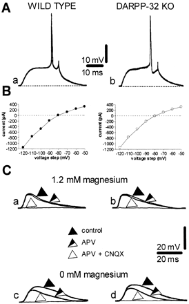
    • No significant difference in the resting membrane potential, input resistance, and current-voltage relationship in neurons recorded from the two groups of animals.
    • AMPA receptor antagonist CNQX suppressed EPSPs in both sets of animals.
      • Removal of Mg$^{2+}$ (removes the voltage dependent block of NMDA receptors) was needed to reveal an NMDA component of the EPSP that could be block by APV (?)
    • High Frequency Stimulation
      • Three spike trains of 100 Hz frequency, 3 s duration with 20 sec ISI of CORTICOSTRIATAL fibers produced 'long-term' chagnes in the AMPLITUDE of EPSPs in wild-type mice.
        • This caused LDP in the presence of Mg$^{2+}$ and LTP in the absence of Mg$^{2+}$
        • When knockout mice were given the same stimulation they show neither LTP nor LDP -> DARPP/PP-1 is necessary for AMPA's LT/DP related activity.
        • Direct test of whether inability to inhibit PP-1 is what causes lack of plasticity
          • Perform the same experiment and then bath the brain in okadaic acid and  calcyculin A (PP-1 inhibitors) 
            • Okadaic acid restores both
            • Calcyculin A restores only LTP
            • No main effect of either on EPSPs
    • Role of D1-like DA receptors and PKA in corticostriatal LTD and LTP
      • Testing the blockade of D1-like DA receptors was effective in block both forms of learning.
        • D1-like DA receptor antagonist SCH 23990 prevented LTD and LTP in wild-type mice.
      • Interesting
        • Next in the cascade is PKA induced phosphorylation of DARPP-32 so let's inhibit that
          • Intracellular injection of H89 (PKA inhibitor) block LTP but not LTD in wild-type mice.
          • However, when this was added to the Mg$^{2+}$ solution both forms of learning were prevented.
          • Suggests different cellular loci for PKA action on learning
            • For LTP but not for LTD this particular pathway is activated POST-synaptically on spiny neurons.
          • More to come...
  • Discussion

Monday, October 31, 2011

Title: Dopamine gating of glutamatergic sensorimotor and incentive motivational input signals to the striatum
Author: Jon C. Horvitz
Journal: Behavioural Brain Research
Year: 2002


  • Overview
    • Traditional assumption: REWARD STIMULI are unique in producing a PHASIC dopamine (DA) response; other stimuli produce much more gradual (necessarily TONIC?) DA responses.
    • Not assumed in this article
    • Forebrain DA activity signals neither the "unpleasantness nor the pleasantness of the event" that elicited it.
    • KEY IDEA: "Activation of substantia nigra (SN) and ventral tegmental area (VTA) DA neurons, and consequent release of nigrostriatal and mesolimbic DA to dorsal and ventral striatal target regions, modulates the processing of concurrent glutamate (Glu) inputs. [This] occurs under conditions of unexpected environmental change."
    • Main question: How does DA transmission in the dorsal and ventral striatum influence behavior?
      • What biological/environmental conditions lead to elevated DA activity?
      • What information is carried by Glu inputs to striatal neurons?
      • How are these Glu inputs modulated by DA activity?
    • What is the ventral striatum?
      • Nucleus accumbens
      • Caudate-putamen
      • Olfactory tubercle
    • Here the ventral striatum means nucleus accumbens.
    • Instead of thinking of DA as a 'chemical code' for reward, here it is argued that "environmentally elicited elevations in mesolimbic and nigrostriatal DA activity gate the input of reward signals to the striatum, just as they do for sensorimotor signals [...]"
      • Such reward signals are likely to originate in orbitofrontal cortex and basolateral amygdala.
    • What this means is that DA 'informs' striatal cells that an unexpected, important event has occurred.
  • DA neurons respond to salient unexpected events
    • It is unlikely that heightened attentional states are associated with a phasic DA response, because attentional systems would likely be recruited by both the presence of an unexpected event and the absence of an expected event. DA neurons increase activity only to the former, while they are inhibited by the latter.
    • Time Course
      • Very rapid onset
        • Phasic activation increase in approximately 50-100 ms after stimulus onset
    • It can't only be REWARD prediction error because of evidence showing that DA in prefrontal cortex (PFC) and nucleus accumbens (NA) is elevated under appetitive and aversive conditions.
    • Primary Aim: "[P]rovide a framework that accounts both for the promiscuous DA response to salient events and for the large body of evidence showing that DA disruptions attentuate the impact of rewards (and punishers) on several aspects of behavior and learning."
    • Important: DA may be considered a gatekeeper of glutamatergic information flow to the striatum.
  • DA selectively promotes the processing of strong glutamate inputs to the striatum
    • It turns out that DA acting at the D1 receptor increases the activity of glutamate at NMDA receptor sites. D2 however, does the opposite and reduces activity of glutamate at non-NMDA receptor sites.
  • DA activity gates the throughput of sensorimotor and incentive motivational inputs to the striatum
    • Striatal neurons receive inputs from a wide variety of 'types' of neurons. For example, the caudate, putamen, and ventral striatum contain neurons that respond to arm movements but only when the animal expects to receive a food reward following the movement. Conversely, very little activity is seen if a sound is expected to follow the movement.
    • Striatal neurons respond to very abstract types of information.
    • Ah, logic is so beautiful: [Individual striatal neurons] ... appear to be capable of representing the conjunction of two conditions[!]
      • Striatal neurons are the (and CONDITIONS ...) Lisp function!
    • What happens when nigrostriatal DA transmission is reduced?
      • Not surprisingly, input signals are less likely to produce a response.
      • Interestingly, when DA is depleted the striatal cell response is diminished but only in background activity NOT phasic activity. When DA is increased the phasic response is stronger than baseline and the background activity is actually reduced!
    • There's evidence that suggests that DA modulates both the striatal response to current glutamate inputs and log-term changes in synaptic strength of these inputs.
  • Striatal plasticity: stimulus-response learning, salience assignment to synaptic inputs, and/or stimulus-response-outcome chunking
    • Schultz thinks that DA signals provide the 'difference' between reward occurrence and reward prediction. "The phasic DA response increases the synaptic strength betwee ncurrently ative striatal input and output elements, increasing the future likelihood that the current set of corticostriatal inputs (reward) will activate striatal outputs (motor responses)." The problem with this hypothesis is that if DA additionally responds to aversive events then modulation of behavior via this mechanism would mean that behavioral responses leading to aversive outcomes would be increased in number. So, says JH, DA's primary function will not likely be one involving the strengthening of stimulus-response (S-R) connections.
    • So what does DA actually do then?
      • Well, maybe strengthening of corticostriatal inputs via DA serves to increase the future salience of an event (or series of events).
      • According to JCH this implies that additional processing of information is performed later in more specialized regions of basal ganglia. This would then imply that something downstream of the striatum is what is determining the reward value of a stimulus; it isn't indicated by the phasic DA response. He proposes frontal regions as the area that performs response selection.
      • It's an indicator for salient environmental change
        • Biologically this means that, due to the processes mentioned earlier, this change will cause an increase in "glutamate signal-to-noise ratios within dorsal and ventral striatal target sites"
  • Thoughts and Questions
    • What does it mean when 'a representation converges' onto a neuron?
      • How do non-physical things (representations) converge onto physical things (neurons)?

Friday, October 28, 2011

Testing out Google's code prettifier...
% a MATLAB function
function x = some_func()
fs = 1000;
d = 1;
t = 0:1 / fs:d;
x = sin(t);
ex = 0.1 * randn(size(x)) + x;
end
class Point(object):
    """
    """

    def __init__(self, x, y):
        """__init__

        Arguments:
        - `x`:
        - `y`:
        """
        self._x = x
        self._y = y

    @property
    def x(self):
        return self._x

    @x.setter
    def x(self, value):
        self._x = value

    @x.deleter
    def x(self):
        del self._x